Today and tomorrow students worked on making their study guides to review for the Cell Cycle/Mitosis and Cancer quiz on Friday.
Here's what you need to know:
All the stages of the cycle cycle, the order and what the stages of mitosis look like
The differences in cytokinesis between plant and animal cells
Information from the cancer packet (the questions you answered)
What angiogenesis is and how it is related to cancer.
In addition to the content, you will be tested on your microscope skills. I will ask you to find one of the stages in Mitosis using slide of plant cells. You will have two attempts to do this successfully. Each attempt can take no more than five minutes. This will count as a pass / fail laboratory grade.
Showing posts with label microscopes. Show all posts
Showing posts with label microscopes. Show all posts
Wednesday, November 30, 2011

Wednesday, October 12, 2011
Microscopes
Quote of the day, "This is so awesome! Have you seen the liver?! It's crazy!"
After completing a binder check C period continued working with microscopes. G period learned about proper microscope use and tomorrow G period will have their binder check.

After completing a binder check C period continued working with microscopes. G period learned about proper microscope use and tomorrow G period will have their binder check.
Wednesday, September 8, 2010
Microscopes
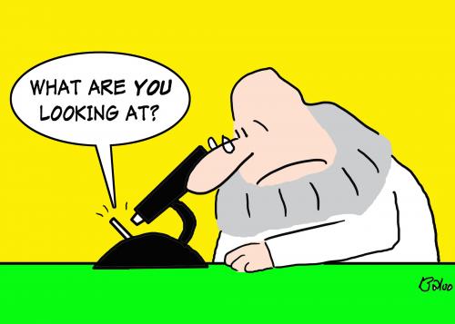
Today we started working with microscopes. After reviewing the parts of the microscope, students practiced looking at different tissue samples, drawing them and viewing the samples under various powers.
Homework: none.
Tuesday, November 3, 2009
Microscopes, Limits to cell size, stages of mitosis
Today students took notes on the three types of microscopes, limits to cell size and the stages of mitosis. 
Homework: Complete "Mitosis Homework #1."

Homework: Complete "Mitosis Homework #1."
Monday, November 2, 2009
Starting Mitosis
Today students examined cells undergoing mitosis.  Here's what happens in the cell:
Homework: Read pages 246-248. Draw the stages of mitosis, in order, with labels (centrioles, nuclear envelope, chromatin, centromere and spindle -- where appropriate).
 Here's what happens in the cell:
Homework: Read pages 246-248. Draw the stages of mitosis, in order, with labels (centrioles, nuclear envelope, chromatin, centromere and spindle -- where appropriate).
Monday, September 14, 2009
Introduction to Microscope Work
Today students had an introduction to working with microscopes which included identifying the parts of the microscope, proper techniques and looking at a variety of slides. Students had the opportunity to look at:
Human blood
Human scalp with hair and sweat glands
Human lung
Frog skin
Large intestine from a mammal
Pollen from a Lily
Spirogyra (a freshwater algae)
Gills from a catshark
A cross section of stem from a Basswood tree.
HOMEWORK: READ THE PROCEDURE FOR LAB 1: MAKING WET MOUNT SLIDES (posted above).
You will have a pre-lab quiz on the procedure. You may use any notes that are in your own handwriting. You do NOT need to print a copy of the lab.
Human blood
Human scalp with hair and sweat glands
Human lung
Frog skin
Large intestine from a mammal
Pollen from a Lily
Spirogyra (a freshwater algae)
Gills from a catshark
A cross section of stem from a Basswood tree.
HOMEWORK: READ THE PROCEDURE FOR LAB 1: MAKING WET MOUNT SLIDES (posted above).
You will have a pre-lab quiz on the procedure. You may use any notes that are in your own handwriting. You do NOT need to print a copy of the lab.
Subscribe to:
Posts (Atom)